
Hip & Groin Conditions
Femoroacetabular Impingement (FAI): Hip Impingement Treatment in Mayfair, London
If you have been given a diagnosis of femoroacetabular impingement and told that surgery is the answer, it is worth pausing before accepting that as settled. The UK’s randomised controlled FAIT trial, the largest clinical study comparing arthroscopic hip surgery with physiotherapy for symptomatic FAI, found that a structured programme of physiotherapy and activity modification produced outcomes comparable to surgery at eight months, with both groups improving meaningfully. Surgery is not wrong for FAI when the clinical picture warrants it. But it should not be the default first step for a condition that responds substantially to well-delivered conservative management.
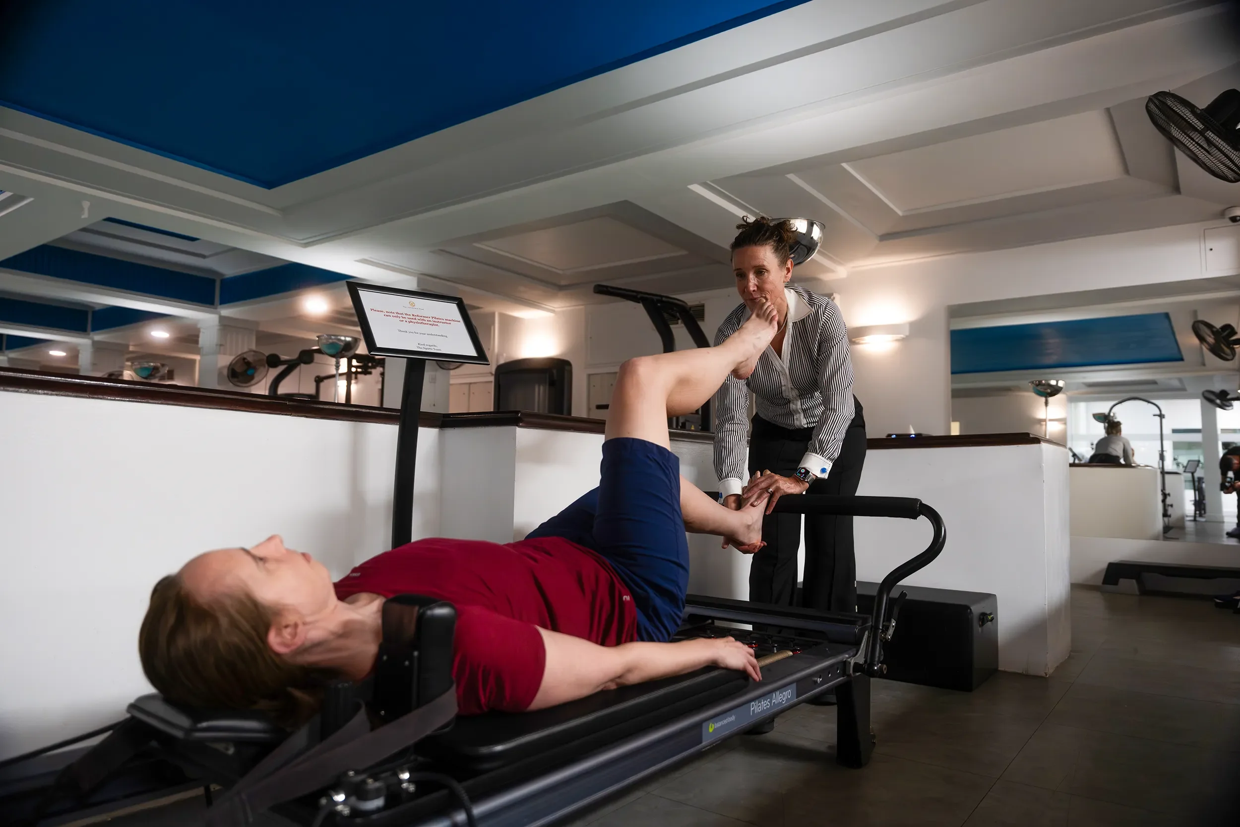
At our Mayfair clinic, we treat femoroacetabular impingement in active professionals, recreational athletes, and competitive sports people across a wide range of disciplines. Our approach begins with a thorough clinical assessment, an honest picture of what conservative management can realistically achieve for your specific hip, and a direct referral when the surgical conversation is the right one.
What Is Femoroacetabular Impingement?
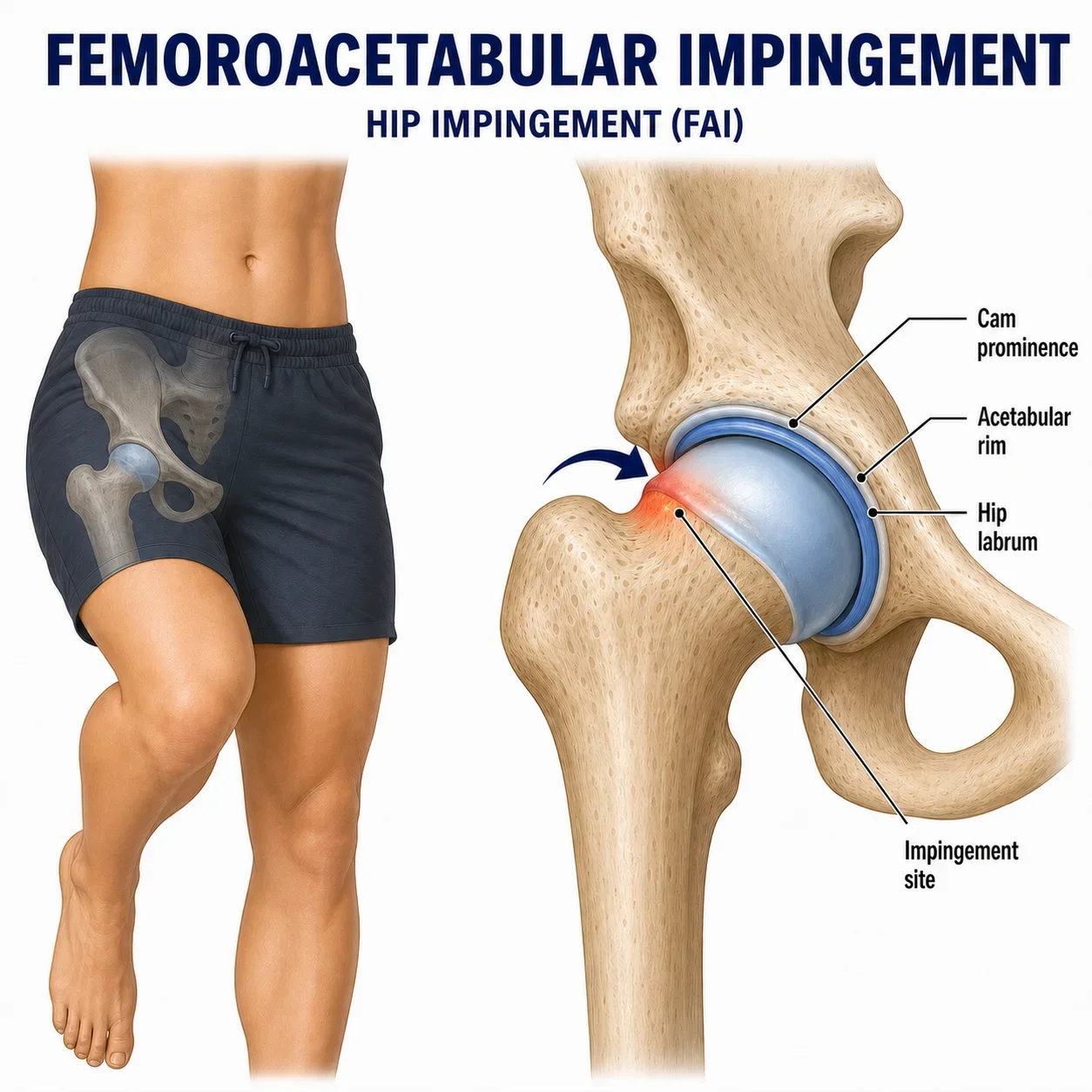
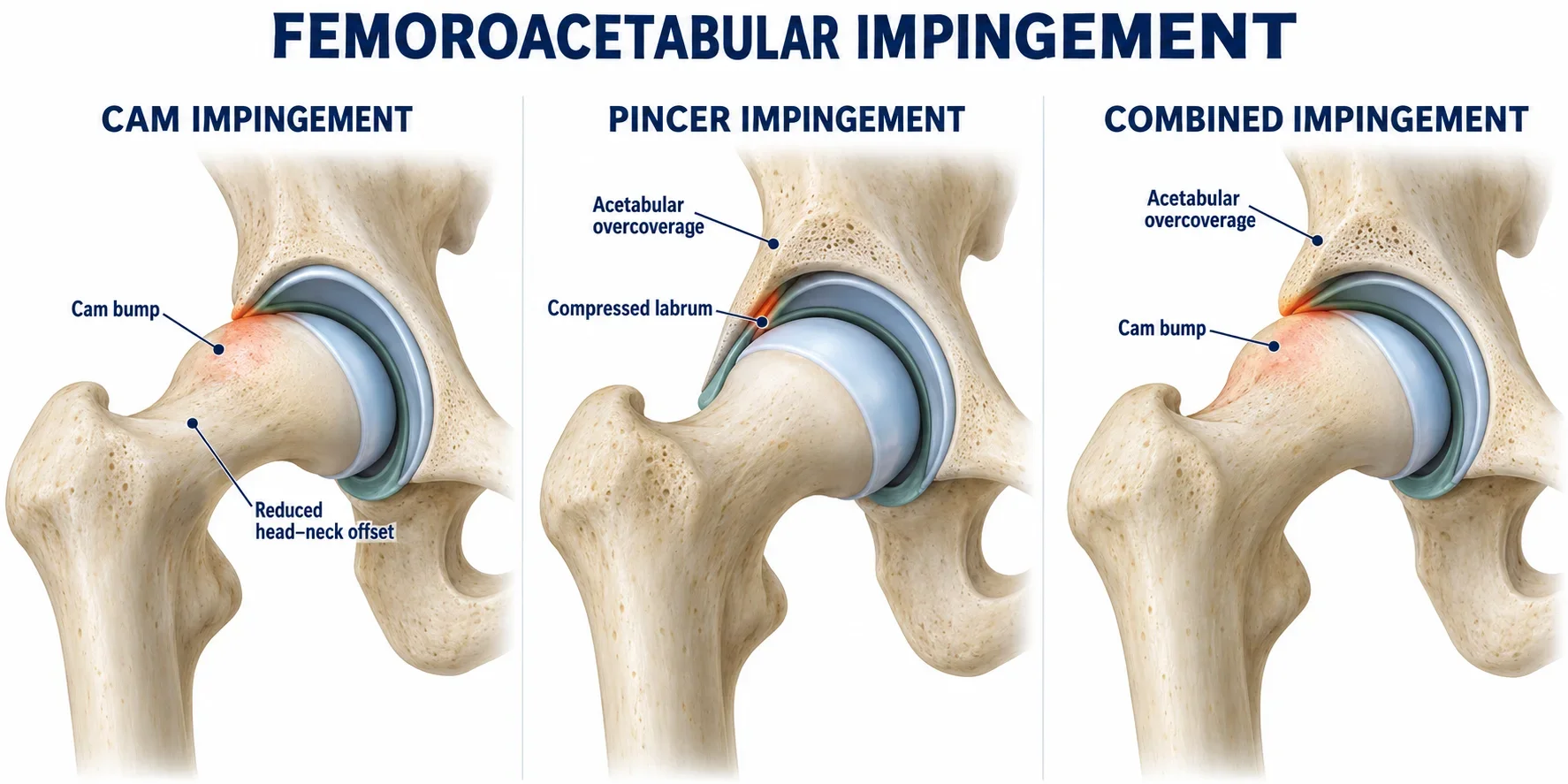
Femoroacetabular impingement is a condition caused by a mismatch in the shapes of the ball and socket of the hip joint. The ball is the femoral head, the rounded top of the thigh bone. The socket is the acetabulum, a cup-shaped structure in the pelvis. In a well-shaped hip, the ball moves freely within the socket without the neck of the femur contacting the rim of the acetabulum. In FAI, a bony abnormality on the ball, the socket, or both causes premature contact between the femoral neck and the acetabular rim during movement, most commonly at end-range hip flexion and internal rotation.
This repeated abnormal contact does not happen once and cause a discrete injury. It happens with every repetition of the provoking movement, over months and years, generating progressive damage to the labrum (the fibrocartilage ring at the entrance to the socket) and eventually to the articular cartilage lining the joint itself. The labral damage is the structural consequence of the impingement: the bony mismatch is the problem. Understanding this distinction matters because it explains why treating the labrum in isolation, without addressing the mechanical environment that is damaging it, produces unreliable results.
The Three Types of FAI
Cam Impingement
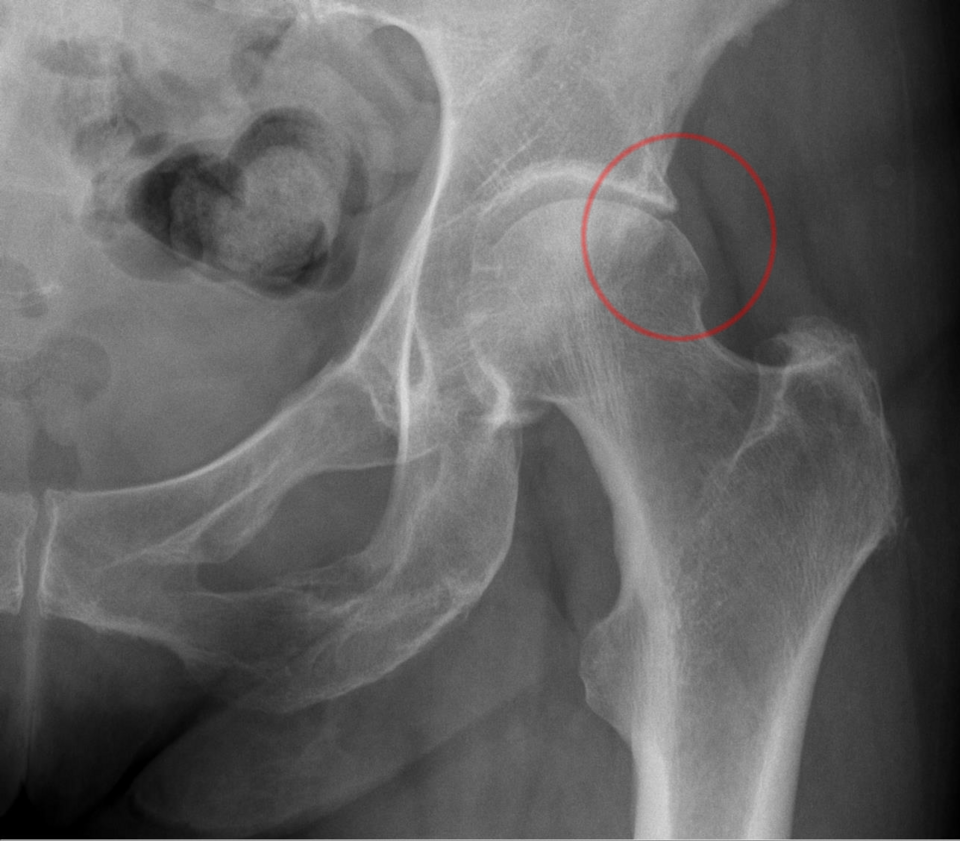
Cam impingement is the most common type and tends to affect young men, though women are also affected. A cam deformity is an abnormal bony prominence at the femoral head-neck junction: the ball is not perfectly round, and this extra bone repeatedly contacts the acetabular rim as the hip flexes. Over time, this creates fraying and tearing of the labrum at the point of contact, followed by delamination of the articular cartilage from the socket wall. If left untreated, the cartilage damage can progress to joint surface loss, which is the beginning of hip osteoarthritis. The severity of the cam lesion is quantified by the alpha angle on MRI: an angle above 55 degrees is generally considered abnormal.
Combined Cam and Pincer Impingement
It is entirely possible to have abnormalities of both the ball and the socket simultaneously, and combined morphology is identified at imaging in a significant proportion of symptomatic FAI patients. The clinical management and surgical approach, when surgery is indicated, account for both components. Combined FAI does not automatically carry a worse prognosis than either type alone, but it does require thorough assessment of which morphology is the primary driver of symptoms.
Pincer Impingement
Pincer impingement is caused by the socket rather than the ball: the acetabulum covers too much of the femoral head, so the labrum is pinched between the femoral neck and the over-prominent acetabular rim with hip flexion. It tends to present in middle-aged women. The prognosis is generally more favourable than cam impingement because pincer-type damage is less likely to progress to the cartilage delamination and osteoarthritis associated with cam lesions. Pincer morphology is identified by the lateral centre-edge angle and acetabular index on plain X-ray and MRI.
Symptoms and Who Is Affected
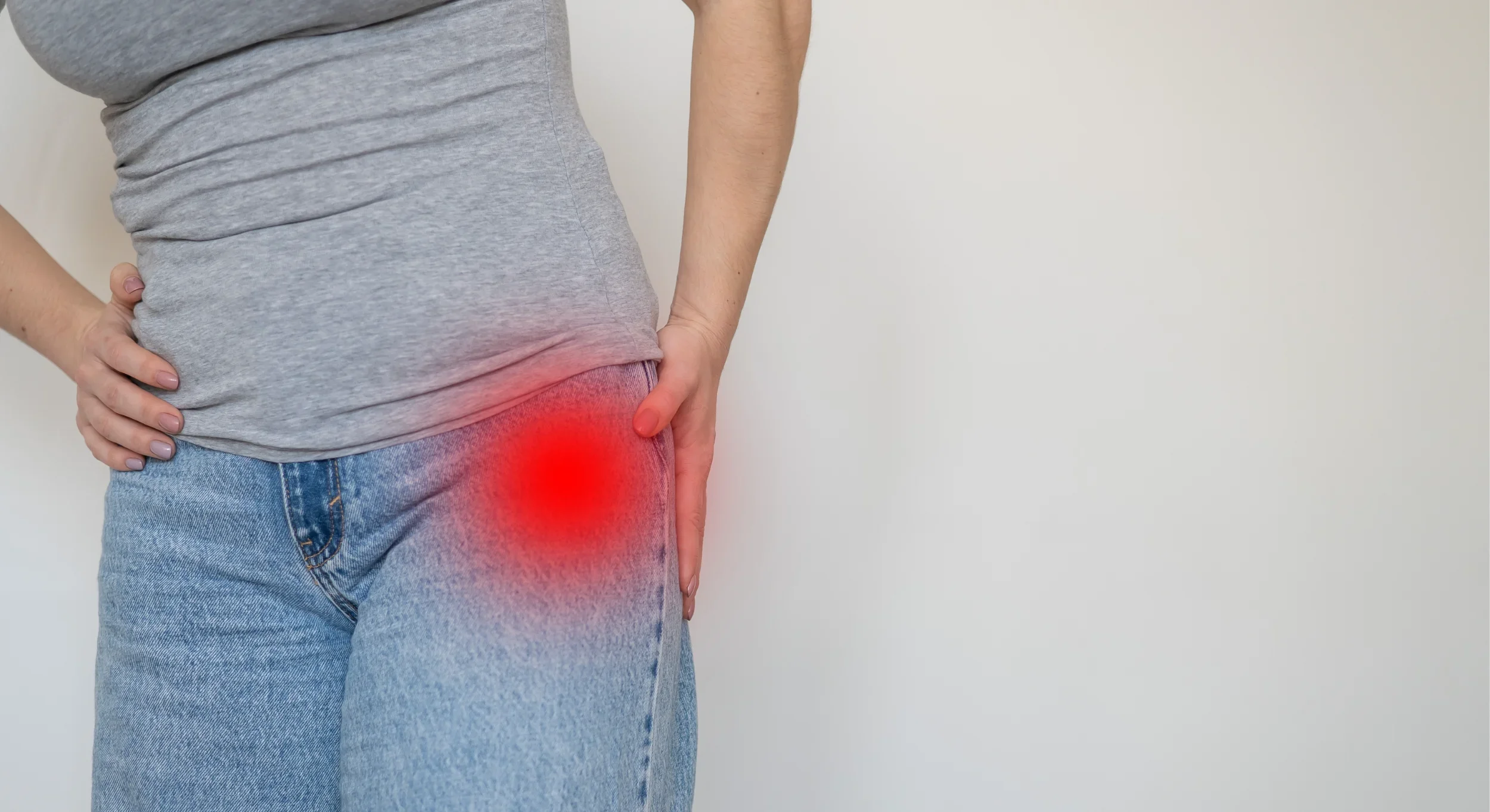
The hallmark of FAI is anterior groin pain: a deep aching discomfort at the front of the hip, often described as sitting at the crease of the groin rather than over the outer hip where patients often point when asked where their hip hurts. Pain may also radiate around the side of the hip in a C-shape. It is typically provoked by end-range hip flexion, sustained sitting, getting in and out of cars, climbing stairs, and rotational movements. A pinching or catching sensation in the groin at the end of hip flexion is characteristic. Many patients report that pain comes on after rather than during activity, particularly after running, cycling, or training sessions.
FAI is most commonly symptomatic in physically active adults between their twenties and forties. The bony morphology develops during adolescent skeletal growth, and sports that demand high-volume hip flexion during this period, including football, rugby, basketball, martial arts, and gymnastics, are associated with higher rates of cam morphology. Cycling in an aggressive position, rowing, and any sport requiring deep hip flexion under load can provoke symptoms in a hip that has been asymptomatic for years. Many patients have had the morphology throughout their adult life and develop symptoms only when training load changes, sitting position changes, or surrounding soft tissue tightness reduces the available joint space.
It is important to note that bony cam and pincer morphology is extremely common in the general population and does not automatically cause symptoms. FAI syndrome requires the combination of bony abnormality, characteristic symptoms, and specific clinical signs. An MRI finding of a cam lesion in the absence of groin pain and a positive clinical examination is not necessarily a condition requiring treatment.
Diagnosis: Clinical Assessment Before Imaging
FAI is a clinical diagnosis supported by imaging, not an imaging finding in the absence of symptoms. The assessment begins with a thorough history and clinical examination. The FADIR test (Flexion, ADduction, Internal Rotation) positions the hip in the posture of maximum impingement: a positive test, where this reproduces the patient’s groin pain, is the most sensitive clinical indicator of FAI. Hip range of movement, particularly internal rotation and hip flexion, is assessed and compared bilaterally. Hip strength, particularly the deep rotators and gluteal muscles, is measured using VALD dynamometry.
When imaging is needed, high-resolution MRI (ideally 3T) is the investigation of choice. It shows the bony morphology, the state of the labrum, the articular cartilage health, and the surrounding soft tissues, while also excluding other conditions that can present similarly, including avascular necrosis and hip stress fractures. Plain X-ray provides useful information about bony shape and joint space but cannot assess soft tissue structures.
Where there is diagnostic uncertainty about whether pain is truly coming from within the hip joint rather than from the surrounding soft tissues or referred from the lumbar spine, a diagnostic intra-articular injection of local anaesthetic can clarify the picture. If the injection produces meaningful relief, this confirms the hip joint as the primary pain source. We have access to sports medicine physicians and musculoskeletal radiologists for guided injections and specialist imaging when these are clinically indicated.
Surgery or Physiotherapy: Making the Right Decision
The question most patients with FAI arrive with is whether they need surgery. The answer depends on what has already been tried, the severity of the morphology and associated damage, and the realistic demands of the patient’s sport or activity. What it should not depend on is the assumption that surgery is automatically superior to conservative management, because the evidence does not support that assumption.
A structured conservative programme that addresses hip rotator and abductor weakness, movement control, and activity modification can produce meaningful and sustained improvement in the majority of patients with symptomatic FAI. The cases where surgery is clearly the right first or next step are those where the bony morphology is severe, significant cartilage damage is already present, conservative management has been given a genuine and structured trial without adequate improvement, or the patient’s activity level and goals are not compatible with the timeline and constraints of conservative management. For a competitive athlete who needs to return to contact sport in a defined timeframe, the surgical and conservative timelines have to be weighed explicitly.
We make this assessment honestly. If we think surgery is likely to be necessary, we say so and refer directly. If we think a structured conservative programme is the right first approach, we commit to making it as effective as possible. We have established relationships with hip arthroscopists in London and can arrange surgical opinion without delay when it is warranted.

Conservative Management at Physio Remedies
Effective physiotherapy for FAI is not a generic hip exercise programme. It is a structured, progressive rehabilitation built around the specific strength and movement control deficits that increase joint contact forces and provoke symptoms.
We begin with objective measurement. VALD dynamometry tests hip rotator and abductor strength bilaterally, quantifying the deficit and establishing a baseline against which progress is tracked. Hip flexor and rotator range of movement is assessed and the positions that provoke impingement are identified. The relationship between lumbopelvic control and hip position under load is examined, because many patients with FAI have movement patterns, both in sport and daily activities, that consistently drive the hip into impingement and that will maintain symptoms regardless of how much strengthening is done around them.
The programme addresses all of this in parallel: progressive strengthening of the deep hip rotators, gluteal muscles, and hip abductors; movement retraining to reduce provocative hip positions during sport, strength training, and daily life; and activity modification to reduce impingement loading during the period when the hip is sensitised. This last point is not rest. It is strategic reduction of the specific movements and loading patterns driving symptoms, while maintaining fitness and overall conditioning. We give explicit guidance on what to modify, what to continue, and how to progress.
After Hip Arthroscopy: Rehabilitation That Matches What Was Done
If arthroscopic surgery is the right decision, the post-operative rehabilitation needs to match the specific procedures performed. Hip arthroscopy for FAI typically involves bony correction of the cam and or pincer lesion, combined with management of the labral damage at the point of impingement. Where the labrum can be repaired it usually is, because a functional labrum maintains the intra-articular fluid seal that protects the cartilage. Where it has degenerated beyond repair, debridement is performed instead. These two procedures have different healing timelines and different early loading constraints, and we read the operative notes before designing the programme.
In the early weeks, the priority is managing post-operative pain and swelling, restoring hip range of movement within the surgeon’s prescribed limits, preventing the significant muscle atrophy that accumulates quickly in this period, and beginning very gentle activation of the hip rotators and abductors. Where the healing tissues limit conventional loading, Blood Flow Restriction training allows meaningful strength stimulus at the low absolute loads that are safe in this phase. As the hip heals and loading constraints are lifted, the programme progresses through progressive strengthening, functional movement retraining, and sport-specific loading.
We track hip rotator and abductor strength throughout using VALD dynamometry, with the 90% symmetry threshold as the benchmark for return-to-sport clearance. VALD ForceDecks provides objective single-leg loading and functional assessment data in the later stages. Return to full sport is confirmed by testing rather than timelines: the patients who return too early, before the repaired tissue has the strength and mechanical environment to tolerate full loading, are the patients who represent with the same problem.
Frequently Asked Questions
Here are answers to some of the common questions we receive regarding FIA at Physio Remedies.
-
Not necessarily as a first step. The FAIT trial evidence shows structured physiotherapy producing comparable outcomes to arthroscopic surgery at eight months. Surgery is appropriate when conservative management has had a genuine structured trial without adequate improvement, when the morphology or cartilage damage is severe, or when the patient’s activity demands make conservative management impractical. We assess which situation you are in and give you a direct recommendation rather than a generic answer.
-
A deep aching pain in the front of the groin, often reproduced by sustained sitting, getting in and out of cars, climbing stairs, and end-range hip flexion. A pinching or catching sensation in the groin is characteristic. Pain frequently comes on after rather than during activity. It is commonly mistaken for a groin strain or tight hip flexors, and many patients have been treated for the wrong condition before FAI is identified.
-
FAI is the mechanical condition: the bony mismatch between ball and socket that causes premature contact within the hip joint. The labral damage is what happens to the soft tissue ring at the entrance to the socket when it is repeatedly compressed and abraded by that contact over time. The labrum is injured as a consequence of the impingement. Treating the labrum without addressing the bony mechanics that damaged it is why some surgical outcomes are incomplete.
-
Usually yes, with modifications. The goal is to identify the specific movements that provoke impingement and reduce those strategically, while maintaining fitness and progressive loading in positions the hip can tolerate. Complete rest is rarely indicated and is often counterproductive. We provide specific guidance on what to modify, what to continue, and how to progress through a structured return to full activity.
-
A structured conservative programme requires three to six months before a meaningful assessment of effectiveness can be made. Some patients improve significantly within eight to twelve weeks. Where meaningful improvement has not occurred by three to four months of well-delivered rehabilitation, the surgical conversation becomes appropriate. The length of post-surgical rehabilitation depends on the procedure: return to competitive sport typically takes five to six months after labral repair and four to five months after debridement alone.
-
No. Cam morphology is found on imaging in a significant proportion of asymptomatic people, particularly those who were active in sport during adolescence. A bony finding on imaging without symptoms and without a positive clinical examination is not FAI syndrome and does not require treatment. Awareness of the activities that provoke FAI symptoms and monitoring is appropriate. If symptoms develop, assessment and a structured management plan at that point is the right response.
-
Quite possibly. An aggressive cycling position, with the hip in sustained flexion and some internal rotation, is one of the most reliably provocative positions for cam FAI. Many people have the morphology for years without symptoms and develop groin pain when cycling position changes, saddle height drops, or training volume increases significantly. Saddle height and cycling position assessment, combined with a hip strengthening programme, is the starting point. If symptoms do not settle with these measures, full FAI assessment is warranted.
-
Not always before starting, but often before committing to a management pathway. Clinical examination alone is sufficient to begin a trial of conservative management in many cases. If symptoms are not improving as expected, if the clinical picture suggests significant cartilage damage or other pathology, or if surgical opinion is being sought, MRI is essential. We can arrange imaging directly when it is clinically indicated.
-
If the bony impingement continues without management, labral damage progresses and articular cartilage delamination follows, particularly with cam-type morphology. This cartilage damage is not reversible and, over time, can lead to joint surface loss and the development of hip osteoarthritis. Not every case of FAI progresses to this outcome, and the timeline varies significantly by morphology type and individual loading, but untreated cam impingement carries a meaningful risk of accelerated joint degeneration.
Book Your Hip Assessment in Mayfair
If you have been diagnosed with femoroacetabular impingement, or if you have deep anterior groin pain that has not been properly assessed, your first appointment lasts one hour. We will examine the hip clinically, review any existing imaging, tell you honestly what conservative management can achieve for your specific presentation, and give you a clear plan.
If you are searching for FAI physiotherapy or hip impingement treatment in London, we are based at the Lansdowne Club in Fitzmaurice Place, Mayfair, a few minutes’ walk from Green Park station.
If you prefer to reach us directly: