
Hand & Wrist Conditions
TFCC Tear Treatment in Mayfair, London
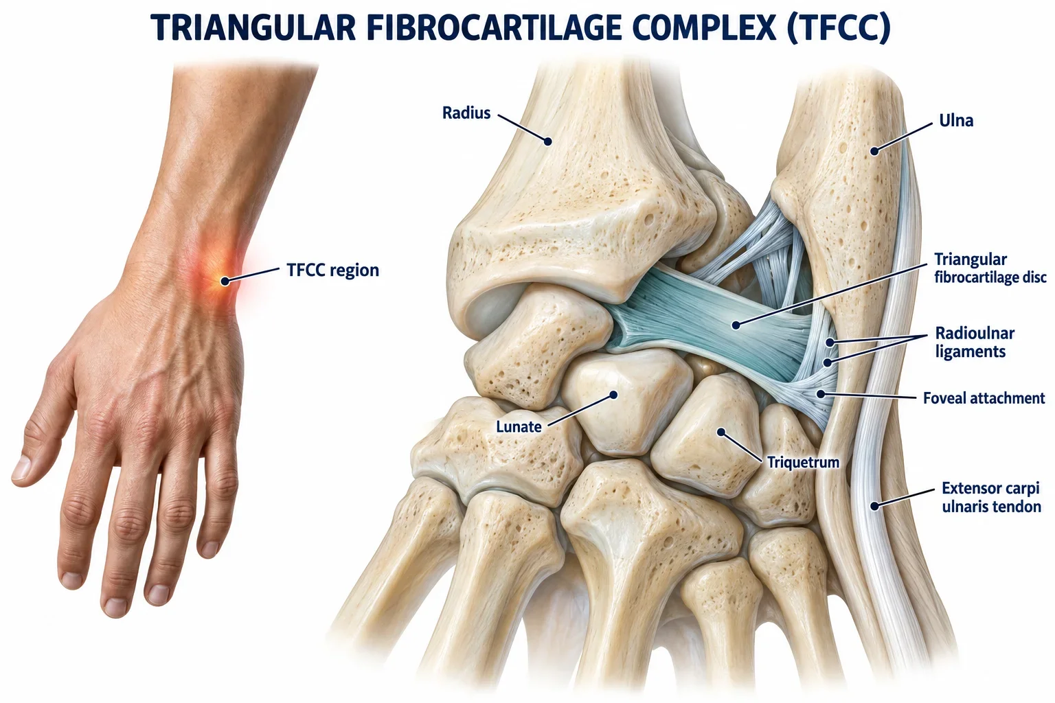
Pain on the little finger side of the wrist is one of the more frequently mismanaged presentations we see in clinic. It is often labelled a wrist sprain, given a support bandage, and left to resolve on its own. Sometimes it does. But when the pain persists, when grip strength has not returned, when there is a click or clunk during rotation that was not there before, the underlying problem is usually more specific: an injury to the triangular fibrocartilage complex, or TFCC.
The TFCC is a structure on the ulnar side of the wrist, towards the little finger, that serves two important functions simultaneously. It acts as a shock absorber between the ulna and the carpal bones, and it provides stability to the distal radioulnar joint, the joint between the radius and ulna just above the wrist that allows the forearm to rotate. When it is injured, whether through a sudden event or through accumulated load, the result is pain with grip, weakness with rotation, and in more significant injuries, a sense of instability or mechanical dysfunction in the wrist.
TFCC injuries are more common than most people realise, and they are treatable. The majority respond well to conservative management when that management is structured correctly. The minority that need surgical input do better when that decision is made promptly. Getting the diagnosis right from the outset determines which path you are on.
Who Gets TFCC Injuries, and Why?
TFCC injuries arrive in two forms: acute, from a single incident, and chronic, from repeated loading over time. The acute injury is typically a fall onto an outstretched hand, or a forced rotation of the wrist under load. The chronic presentation develops more insidiously, with pain and grip weakness building gradually in people whose activities place repeated torsional or compressive stress through the ulnar wrist.
The sporting populations we see most often with TFCC injuries at our Mayfair clinic reflect the range of mechanisms involved:
Tennis players are a consistent TFCC injury population.
The wrist moves through a large range during groundstrokes and serves, and the torque generated through the ulnar side at contact is substantial. Topspin groundstrokes and kick serves in particular load the TFCC in rotation. Players who have developed a technique that relies heavily on wrist snap rather than body rotation through the shot tend to accumulate more load at the wrist over time.
Combat athletes, including boxers and those training in MMA, load the TFCC through specific shot types.
Uppercuts and hooks, and particularly bent-arm shots where the elbow is flexed and the force cannot be efficiently distributed through a straight limb, concentrate stress through the ulnar wrist. Grapplers face a different mechanism: the wrist being caught in an awkward position whilst rolling, or the sustained torsional loading that comes from fighting for grip on a Gi. That fabric grip involves repeated, forceful twisting of the wrist against resistance, and the TFCC is precisely the structure bearing that torsional stress.
Overhead pressing athletes, including those in CrossFit and weightlifting, are at particular risk when shoulder mobility is insufficient.
A clean and press performed without adequate shoulder external rotation forces compensatory movement further down the chain. Load that should be absorbed at the shoulder instead travels through the elbow and wrist, and the TFCC takes more than its share. Assessment of these injuries always involves an in-depth assessment of the strength and mobility of the proximal joints for a better understanding of the mechanisms driving the dysfunction.
Beyond sport, TFCC injuries occur in those who have sustained a wrist fracture, particularly a distal radius fracture, where the anatomy of the joint can be altered by the injury itself. Degenerative changes also occur with age, and it is worth noting that TFCC abnormalities are found on imaging in a significant proportion of people without any pain or functional loss at all. That matters enormously when interpreting scan results.

Recognising a TFCC Injury: Symptoms and Clinical Signs
The defining feature is pain on the ulnar side of the wrist: the little finger side, at or just below the bony prominence of the ulna. It is typically worse during grip, during forearm rotation, and during any loaded movement that takes the wrist into ulnar deviation. Pressing firmly over the ulnar side of the wrist usually reproduces the pain directly.
In more significant injuries, or those that have been present for some time, there may be a palpable or audible click or clunk during wrist rotation. This catching or clunking, particularly when it represents a change from how the wrist previously moved, is a more significant clinical sign and one that influences our decision about imaging. A loss of grip strength that feels disproportionate to the degree of pain, or a sense that the wrist is unstable or giving way under load, also raises the clinical threshold.
What we are establishing at assessment is whether the TFCC is injured in isolation, whether the distal radioulnar joint has lost its normal stability, and whether there are contributing factors upstream in the forearm, elbow, or shoulder that are either causing the problem or perpetuating it.
When Does a TFCC Injury Need a Scan?
This is a question we are frequently asked, and the honest answer is: less often than many patients expect, but sooner than most clinicians act when it does.
For the majority of TFCC injuries, particularly those without instability, clinical assessment is sufficient to guide management. We do not routinely send every wrist pain for MRI. The reason for that restraint is clinically important: degenerative changes and partial tears within the TFCC are found on imaging in a very large proportion of people who have no pain and no functional loss at all. A scan that reveals a TFCC abnormality is therefore not, on its own, a diagnosis. The imaging findings have to be interpreted alongside the clinical picture.
The situations in which we do move promptly to imaging are specific. A new, painful clicking or clunking during rotation that was not there before the injury. A significant loss of grip strength with a sense of wrist instability or giving way. A presentation that has not responded appropriately to a properly structured trial of conservative management. In these situations, MRI or MR arthrography (MRA) is the imaging modality of choice, providing the most accurate assessment of the degree of TFCC disruption and, critically, whether the distal radioulnar joint remains congruent.
That last point, bony congruency at the distal radioulnar joint, matters for one specific reason. If there is structural disruption combined with instability that is not resolving, and if surgery becomes the appropriate next step, the evidence suggests that outcomes are better when repair is performed within the first seven months of injury, before the surrounding tissues degrade further. We do not wait indefinitely before escalating. When the clinical picture points towards surgical assessment, we act on that promptly.
How We Treat TFCC Injuries
For stable TFCC injuries, which represent the majority of what we see, conservative management is highly effective when it is structured around the right sequence of protection, loading, and progressive return to activity.
The first decision is how much protection the wrist needs. In presentations with mild to moderate pain and no instability, a short period of bracing, typically two to three weeks, reduces the torsional and compressive stress on the TFCC enough to allow the acute irritability to settle. Where pain is more significant, we may extend that bracing period to four to six weeks. The brace does not need to be worn at all times throughout that period; we advise specifically on when it is and is not necessary, because immobilising the wrist entirely for six weeks is not the goal.
What makes our approach different is that we do not wait for bracing to finish before beginning strengthening. Even within a period of relative protection, there are loading strategies that build the muscles supporting the TFCC without generating shear force through the distal radioulnar joint. Isometric work targeting the pronator quadratus, extensor carpi ulnaris, brachioradialis, and triceps can begin early, in positions that respect the injury.
One position that is particularly useful in early TFCC rehabilitation is the hammer curl. With the thumb pointing upward and the palm facing inward, the forearm is held in a neutral position between full pronation and supination. This orientation places significantly less shear stress through the distal radioulnar joint than exercises performed with the forearm fully rotated, and it allows meaningful loading of the supporting musculature at a stage where full rotation would be provocative. It is a clinically useful position that most standard wrist rehabilitation programmes do not use.
As pain settles and the wrist can tolerate more load, we progress to exercises in greater ranges of forearm rotation, gradually reintroducing the movements that reproduce the demands of the patient's sport or activity. For the tennis player, that means working towards the rotational loading of a groundstroke. For the boxer, it means recovery the absorb and generate force through bent-arm punches and maintaining a stable wrist position. For the overhead athlete, it may mean addressing strength or mobility deficits at the shoulder or elbow that are contributing to the wrist being overloaded in the first place.
Return to full sport or activity is milestone-based rather than time-based. Grip strength symmetry, pain-free rotation through full range, and confident loading through the provocative movements of the specific sport are the markers we work towards. For most stable TFCC injuries managed from an early stage, return to full function within twelve to sixteen weeks is a realistic expectation.
When Is Surgery Required for a TFCC Injury?
Surgery is not the outcome for most TFCC injuries. Where there is a clear structural disruption with loss of distal radioulnar joint congruency, where instability persists despite a thorough trial of conservative management, or where the clinical picture warrants it in a competitive athlete, surgical assessment is appropriate. Wrist arthroscopy is the current gold standard, offering both precise diagnosis and the option of simultaneous repair in a single procedure.
When we reach the point of surgical referral, we do so with a direct pathway to hand and upper limb surgeons experienced in wrist pathology. The evidence supporting earlier surgical repair, within the first seven months of injury for acute traumatic tears, informs how quickly we move when it is the right decision. After a successful repair, structured physiotherapy is essential to restore range of movement, rebuild wrist stability, and return the patient to the demands of their sport. The surgical result alone does not do that work.
During the post-operative period, rehabilitation should focus not only on protecting the healing TFCC but also on maintaining the capacity of the proximal kinetic chain, particularly the shoulder and elbow stabilisers. Contralateral training of the unaffected upper limb can also be utilised to minimise deconditioning and preserve neuromuscular function during periods of restricted wrist loading.
Frequently Asked Questions
Here are answers to some of the common questions we receive regarding TFCC treatment at Physio Remedies.
-
Not quite. A wrist sprain is a general term for ligament injury around the wrist. A TFCC injury specifically involves the triangular fibrocartilage complex on the ulnar side, and it affects both the cartilaginous disc and the surrounding ligaments that stabilise the distal radioulnar joint. The management, particularly the question of bracing, loading strategy, and imaging threshold, is more specific than for a general wrist sprain.
-
No. Most TFCC injuries can be assessed and initial management begun on clinical grounds alone. If you have already had imaging, bring the results: they are useful context. If we feel imaging is indicated based on what we find at assessment, we will arrange it through our clinical network rather than asking you to pursue it yourself.
-
Not necessarily. TFCC abnormalities, including partial tears, are found on imaging in a large proportion of people with entirely normal wrist function and no pain. The imaging findings need to be interpreted in the context of your symptoms and clinical examination. If your wrist is functioning well, a scan finding alone is not a reason for intervention.
-
In most cases, yes, with modification. The specific movements to avoid depend on your sport and the nature of the injury. Tennis players may need to modify stroke mechanics temporarily. Boxers may need to avoid certain shot types while the wrist settles. Grapplers may need to avoid the torsional loading of gi work during early management. The goal is always to keep you as active as possible within safe limits, rather than stopping everything.
-
For stable injuries managed conservatively from an early stage, return to full sport within twelve to sixteen weeks is realistic. More established or complex injuries take longer. Where surgery is required, return to full function typically takes four to six months from the date of the procedure, with good outcomes for the majority of patients who follow a proper post-operative rehabilitation programme.
-
We accept Bupa, AXA, and Cigna International. Our initial assessment is £190 for one hour. Please contact us to confirm your cover before your appointment.
Book Your TFCC Assessment at Physio Remedies, Mayfair
If ulnar wrist pain is affecting your grip, your training, or your ability to compete, come and see us. We are based at the Lansdowne Club in Mayfair, a short walk from Green Park and Berkeley Square.
If you prefer to reach us directly: