

Foot & Ankle Physiotherapy
Foot & Ankle Fracture Rehabilitation in Mayfair, London
There is a particular moment that most patients with ankle and foot fractures experience. They attend their final fracture clinic appointment. The X-ray is reviewed. The surgeon says the bone looks good. They are discharged from orthopaedic care. They leave the building and stand on the pavement outside and wonder why they still cannot walk properly.
The bone being healed and the ankle functioning well are not the same thing. A fracture clinic measures bone consolidation. It does not measure calf strength, ankle mobility, proprioception, or the confidence to load the joint under real-world demands. These are the things that determine whether you can walk without pain, manage a flight of stairs, get back to tennis, or trust your ankle in the moment that counts. They do not restore themselves. They require rehabilitation.
Jane Baker, one of our specialist foot and ankle physiotherapists and a member of the Association of Foot and Ankle Physiotherapists, knows this from both sides. She rehabilitated from an ankle fracture herself. She understands what the hospital system provides and what it does not, and what the weeks and months after discharge actually demand. At Physio Remedies in Mayfair, we pick up where orthopaedic care leaves off, and we do it with a level of objective testing and structured progression that most patients have not encountered in their recovery so far.

What the Hospital Gave You and What You Still Need
The orthopaedic system is designed to fix the bone. It does this exceptionally well. The surgery, the hardware, the immobilisation protocol, the fracture clinic follow-up: all of these are oriented toward one outcome, which is bone healing. Once that is confirmed, the system's job is complete.
What the system is not set up to address is everything that happened to the soft tissues, the muscles, and the nervous system while the bone was healing. Six to twelve weeks in a boot or cast causes calf muscle atrophy that can reduce muscle volume by twenty per cent or more. The ankle joint capsule stiffens. Ankle dorsiflexion is lost. The sensory nerve endings within the joint that govern proprioception, your ankle’s ability to sense its own position and trigger protective responses, are disrupted by the injury and by the period of immobilisation. Surgical incisions create scar tissue that tethers soft tissue and restricts joint movement. Gait mechanics change to protect the injured limb, and those compensatory patterns persist long after they are needed.
None of these are measured at a fracture clinic. None of them appear on an X-ray. And none of them resolve simply because you have been told the bone is healed and to start walking normally again. This is not a criticism of the orthopaedic team. It is an accurate description of two different phases of recovery, managed by different disciplines with different tools. The fracture clinic phase is over. The rehabilitation phase is where function is actually restored.
What We Measure That the Hospital Does Not
The first thing we do at your assessment is establish where you actually are, measured objectively rather than estimated from how things feel. Calf strength is tested using VALD hand-held dynamometry against the uninjured side. The deficit is often striking: patients who have been walking for weeks frequently measure at fifty to sixty per cent of their uninjured limb, having had no idea the discrepancy was that large. The 90% symmetry threshold is our benchmark before return to running or sport. The number tells us how much ground has been lost, which direction the programme needs to go, and how quickly it is reasonable to expect progress.
Ankle dorsiflexion range is measured and compared bilaterally. Loss of dorsiflexion after a fracture alters every step taken: the foot compensates by pronating earlier, the knee absorbs forces it should not, and the calf works inefficiently. Manual therapy to restore this range is not optional in fracture rehabilitation. It is the prerequisite for everything else working properly.
Single-leg balance and proprioceptive function are assessed qualitatively and with VALD ForceDecks, which measures the forces and symmetry of loading during single-leg tasks. This gives us the data we need to progress neuromuscular rehabilitation intelligently and to confirm impact readiness before running or sport resumes. Scar mobility is assessed post-surgically: tethered scar tissue over the lateral or medial malleolus is a consistent and consistently underaddressed source of persistent pain and restricted movement.
Fractures We Rehabilitate
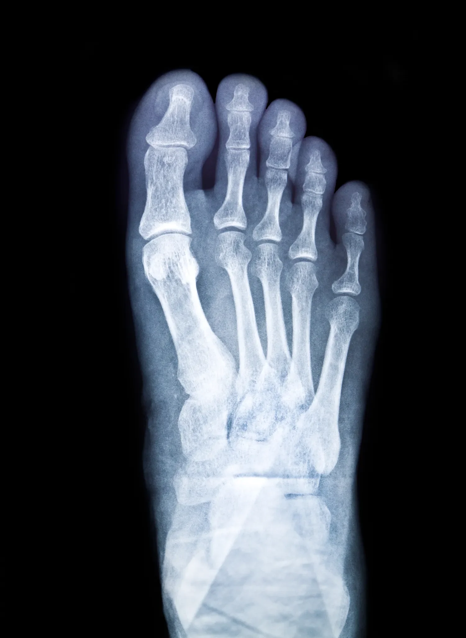
We manage rehabilitation after the full range of traumatic foot and ankle fractures. Ankle fractures, whether a lateral malleolus fracture managed conservatively in a boot or a bimalleolar or trimalleolar fracture requiring surgical fixation with plates and screws (ORIF), are the most common presentation. Calcaneal fractures, typically from falls from height or axial loading, are more complex and have longer timelines: the subtalar joint is often involved and its long-term function is frequently compromised even after good surgical management. Metatarsal fractures, including fifth metatarsal avulsion fractures and more significant shaft fractures, require careful restoration of forefoot loading mechanics alongside the standard strength and proprioception work.
Two fractures warrant specific mention because they are the ones most commonly mismanaged before they reach us. Lisfranc fracture-dislocations are consistently misidentified as midfoot sprains at the time of injury, appear near-normal on standard X-ray, and are only confirmed on weight-bearing imaging or MRI. A Lisfranc injury treated as a sprain is on a reliable path to chronic midfoot instability and post-traumatic arthritis. If you had a significant midfoot injury and persistent midfoot pain, and you have not been specifically investigated for Lisfranc disruption, this needs to be addressed. Talus fractures are rare but carry the specific risk of avascular necrosis from disruption of the bone’s blood supply: the rehabilitation timeline and loading decisions are made in close collaboration with the treating surgeon and are more conservative than for other fracture types for this reason.
How We Rehabilitate Foot and Ankle Fractures
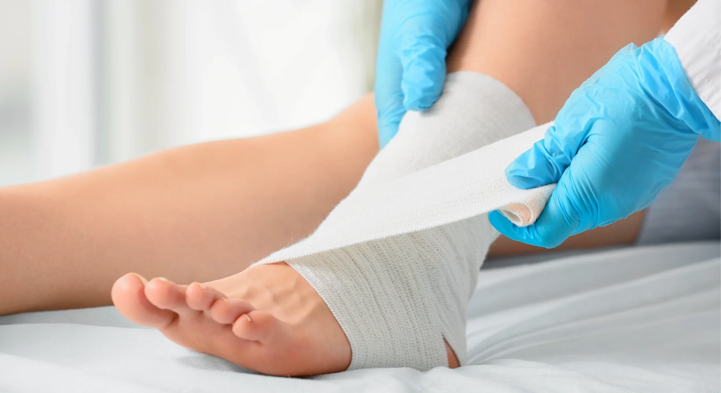
We begin with what is clinically possible at the stage you are at. For patients still in the protected weight-bearing phase, this means non-impact work: joint mobility within the surgeon’s permitted range, foot and calf activation through positions that do not load the fracture site, hip and core work to maintain the fitness that will be needed when loading resumes, and scar management post-surgically. Blood Flow Restriction training is a tool we use extensively during this phase: it applies a cuff to the upper thigh that creates a controlled restriction of venous outflow, allowing the calf and lower limb muscles to be meaningfully loaded at very low absolute weights. The result is genuine hypertrophic stimulus, real strength maintenance, during a period when the limb cannot be loaded normally. It is one of the most clinically significant advances in fracture rehabilitation and it is used by very few physiotherapy practices in London.
Once weight-bearing is permitted and progressive, the programme moves through a structured sequence determined not by the calendar but by what each stage of testing shows. Walking mechanics are addressed before running mechanics. Calf strength is rebuilt on the flat before inclines are added. Single-leg loading is introduced once bilateral loading is solid. Running is not introduced until strength testing confirms the calf is within twenty per cent of the uninjured side and single-leg balance is within normal limits. Return to sport, cutting, jumping, and high-demand loading require 90% strength symmetry confirmed on VALD testing and hop test performance within acceptable limits on VALD ForceDecks. These are not arbitrary thresholds: they are the evidence-based standards for safe return to activity after lower limb injury, and they are what we hold ourselves and our patients to.
Manual therapy runs alongside the loading programme throughout. Ankle joint mobilisation to restore dorsiflexion is a priority from the first weight-bearing session. Scar tissue mobilisation post-ORIF prevents the soft tissue tethering that restricts movement and causes persistent pain months after the wound has closed. Subtalar and midfoot joint work restores the mobility that immobilisation has taken from the joints adjacent to the fracture site. These are not passive treatments added for patient comfort: they are mechanical prerequisites for the loading programme to work effectively.
Working With Your Surgical Team
We read your operative notes and your fracture clinic letters. We understand what hardware is in situ, what the surgical approach was, and what protocol the surgeon has set. We work within that protocol and we contact the surgeon directly if we identify anything during rehabilitation that warrants review: pain that does not fit the expected pattern, hardware that is becoming symptomatic, a Lisfranc rehabilitation that is not progressing as it should, or any sign of post-traumatic arthritic change developing. The gap between orthopaedic care and physiotherapy is not a handover: it is a collaboration. We treat it as one.
We accept Bupa, AXA, and Cigna International insurance. We can arrange imaging, including MRI and X-ray, without a GP referral when it is needed during the rehabilitation process.

“Having broken my ankle, minor break but nonetheless required physio, I was lucky enough to be recommended to Physio Remedies. Sarah Lawson was fantastic and really progressed my rehab so I could get running again which is my passion. Not only that, we worked on my foot and ankle positioning and flexibility which has paid huge dividends beyond just getting me back to full fitness. Highly recommended.”
Frequently Asked Questions
Here are answers to some of the common questions we receive regarding foot and ankle fracture treatment at Physio Remedies.
-
Because the fracture healing and the function recovery are measured differently and happen at different rates. The X-ray shows the bone. It does not show calf strength, ankle mobility, proprioception, or the compensatory movement patterns that have developed over weeks of protecting the injury. These are the things that determine how the ankle actually performs, and they require specific rehabilitation to restore. A clean X-ray is the beginning of recovery, not the end.
-
Yes. The muscle atrophy, joint stiffness, proprioceptive deficit, and altered gait mechanics that accumulate during immobilisation do not resolve on their own. Patients who do not have structured post-fracture rehabilitation consistently have worse long-term outcomes, including persistent pain, chronic swelling, reduced range of motion, and higher rates of subsequent ankle injury.
-
As soon as you are discharged from hospital, and in many cases before weight-bearing is permitted. Non-weight-bearing physiotherapy including joint mobility, non-impact muscle activation, Blood Flow Restriction training, and scar management can begin in the early protected phase and makes a meaningful difference to how quickly the loading programme progresses once weight-bearing resumes.
-
The bone typically consolidates within six to twelve weeks depending on the fracture and whether surgery was required. Walking normally without significant pain or swelling usually takes three to six months. Return to running or sport takes longer and is governed by objective strength and function testing rather than a fixed timeline. For complex fractures including calcaneal and Lisfranc injuries, timelines are longer and the eventual functional endpoint depends on the fracture severity.
-
Persistent swelling after a healed ankle fracture reflects ongoing soft tissue inflammation, altered fluid dynamics, and often the demands being placed on an ankle that does not yet have the calf strength to handle them efficiently. As strength is restored and gait mechanics normalise, swelling typically improves. If swelling is increasing or associated with warmth and pain, it warrants clinical review.
-
The Lisfranc joint complex is the system of bones and ligaments connecting the forefoot to the midfoot. Lisfranc injuries are the most consistently misdiagnosed fractures in the foot: they appear near-normal on standard X-ray, mimic a midfoot sprain in presentation, and are only confirmed on weight-bearing imaging or MRI. If you had a significant twisting or crushing injury to the foot and have persistent midfoot pain or structural asymmetry, Lisfranc disruption needs to be specifically excluded. Treating it as a sprain produces reliably poor outcomes.
-
When your calf strength has reached 90% of the uninjured side on VALD testing, your single-leg balance and hop performance are within normal limits on VALD ForceDecks assessment, and you can walk on all surfaces without pain or significant compensatory movement. For most surgically managed ankle fractures, this is six to nine months from injury. We test rather than estimate.
-
This is set by your orthopaedic surgeon based on the fracture type and surgical approach. Most ankle fractures involve six weeks of non-weight-bearing followed by a graduated loading protocol. Complex fractures, particularly calcaneal and Lisfranc injuries, require longer protected periods. We work within whatever protocol your surgeon has prescribed.
-
Yes, and scar management is a routine part of post-ORIF rehabilitation. Tethered scar tissue over the incision site restricts soft tissue mobility, limits ankle movement, and is a consistent source of persistent pain months after surgery. Manual scar mobilisation, begun once the wound is fully healed, reduces tethering and improves both movement and comfort. This is something that is almost never addressed in standard post-fracture physiotherapy.Item description
-
Blood Flow Restriction (BFR) training uses a cuff around the upper thigh to create controlled restriction of venous return, allowing meaningful muscle stimulus at very low loads. During the non-weight-bearing phase of fracture rehabilitation, it allows calf and lower limb strengthening to continue at loads that are safe for the healing bone. It significantly reduces the muscle atrophy that would otherwise accumulate before full loading is permitted, and it shortens the strength rebuilding phase that follows. We use it regularly in fracture rehabilitation and it is available to very few patients through standard NHS or generic private physiotherapy.
Book Your Fracture Rehabilitation Assessment in Mayfair
Whether you have just been discharged from orthopaedic care, are partway through a recovery that has plateaued, or are aiming to return to sport and want to know objectively where you stand, your first appointment lasts one hour. We will measure what has been lost, tell you honestly what needs to happen, and give you a clear plan with milestones rather than timelines. The assessment costs £190.
If you are searching for ankle fracture physiotherapy or foot fracture rehabilitation in London, we are based at the Lansdowne Club in Fitzmaurice Place, Mayfair, a few minutes’ walk from Green Park station.
If you prefer to reach us directly: